
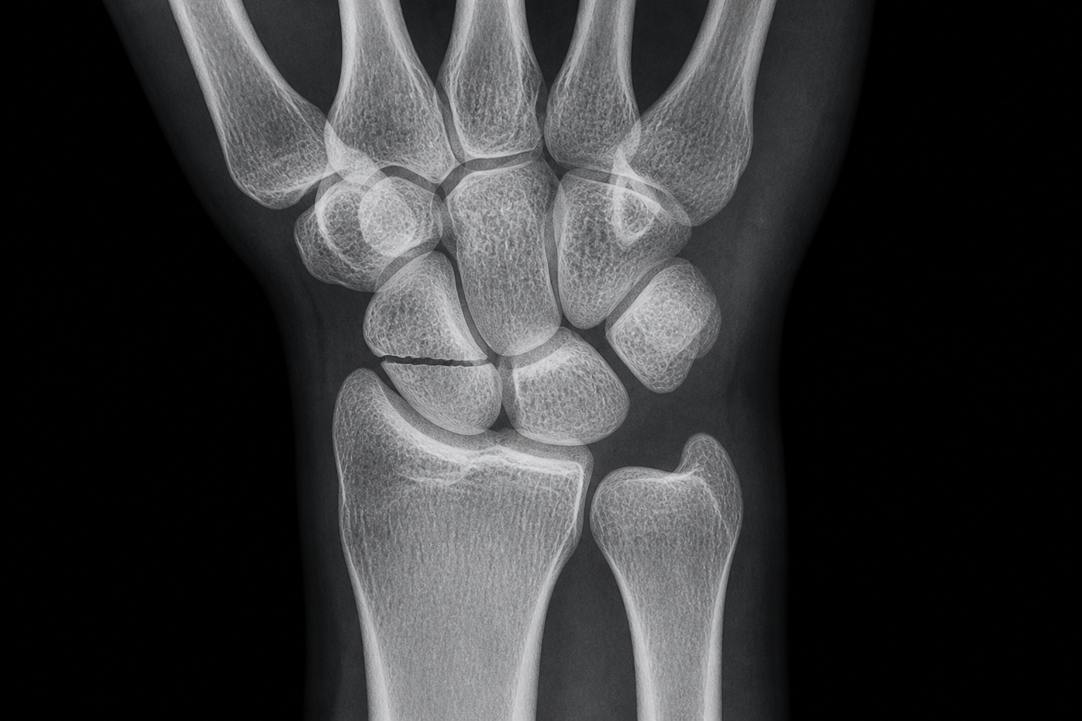
SNAC is short for scaphoid nonunion advanced collapse. It describes a recognised pattern of wrist arthritis that develops in people who have a scaphoid fracture that has never healed. Many of these injuries happened decades earlier and were dismissed at the time as a sprain, often without medical attention being sought at all. The fracture itself may cause little or no trouble for years. What eventually brings a patient to clinic is not the old fracture, but the arthritis that develops afterwards, following a specific and well-documented sequence through the wrist.
This article explains what SNAC actually is, why it happens, how it is staged, and what treatment looks like at each stage, from the least invasive options through to the operations reserved for the most advanced cases.
A scaphoid nonunion means a fracture in the scaphoid bone has failed to heal, leaving two or more fragments that never join back together. The scaphoid sits at the base of the thumb and forms an intricate mechanical relationship with the other bones of the wrist, so an unhealed fracture there produces pain every time weight is put through the hand, a fist is made, or something is lifted.
To understand why, it helps to know how the wrist is built. The forearm has two bones, the radius and the ulna. Below that sit eight small carpal bones. Three of these, the scaphoid, lunate, and triquetrum, work together as a single unit, known in medical terms as an intercalated segment. This is the link between the bones of the forearm and the bones of the hand, and it lets forces pass smoothly from one to the other. The scaphoid has a natural tendency to flex forward, because of how it is shaped and constrained where it meets the trapezium and trapezoid. The triquetrum has an opposite tendency to extend backward, constrained by the hamate. In an intact wrist, these two opposing tendencies stay balanced because both bones are anchored to the lunate by strong ligaments.
When the scaphoid fractures and fails to heal, this balance breaks down. The fragment still attached to the lunate follows the lunate into extension. The fragment nearer the fingertips, no longer held by that connection, flexes on its own. The result is that the scaphoid-lunate-triquetrum unit splits into two independently moving parts instead of one. That abnormal movement gradually wears away the smooth cartilage lining the joint, in a pattern shown to progress in a consistent, predictable sequence.
Degenerative change in an unhealed scaphoid fracture was first formally described in a study of 64 patients with symptomatic scaphoid nonunions, which found arthritic change developing in a consistent order.¹ That sequence is now described in four stages.
Stage I: arthritis develops in the joint between the scaphoid fragment closest to the wrist and the radial styloid, the pointed part of the radius on the thumb side of the wrist.
Stage II: arthritis spreads to the joint between the scaphoid and the capitate, the next carpal bone along.
Stage III: arthritis spreads across the entire joint between the capitate and the lunate.
Stage IV: arthritis reaches the joint between the lunate and the radius itself. This stage is comparatively unusual.
Source: Vender MI, Watson HK, Wiener BD, Black DM. "Degenerative change in symptomatic scaphoid nonunion." Journal of Hand Surgery (American Volume). 1987;12(4):514–519. In this study, 75 percent of patients with a nonunion of four years' duration had changes at the joint nearest the wrist, and 60 percent of those with a nonunion of nine years' duration had changes at the midcarpal joint. Figures reflect this published cohort and are not a guarantee for any individual case.
Presentation varies enormously. The extent of arthritis on an X-ray does not reliably predict how much pain someone experiences. Some patients tolerate SNAC without ever seeking treatment. Others present because a fall brings them to an emergency department, and SNAC is found incidentally on an X-ray taken for the new injury. Others are referred with symptoms that look like carpal tunnel syndrome, and a wrist X-ray taken because the wrist itself feels stiff reveals SNAC as an unrelated finding.
For patients who are troubled by the condition directly, the characteristic story is a continuous, low-grade aching that disrupts sleep and daily function, alongside swelling over the back of the wrist, a noticeable loss of strength, and a restricted range of movement. A common pattern is a patient who has managed activities such as DIY or gardening for years without difficulty, but has noticed that after ten or fifteen minutes they now need to stop and rest the wrist, something they had never needed to do before.
Many patients cannot recall a specific injury at all. This is common in people with physically demanding jobs who injure their hands regularly, and in people whose original injury, decades earlier, was simply dismissed as a sprain at the time.
Once arthritis has developed, SNAC behaves differently from an acute problem such as a trapped nerve. With a trapped nerve, the longer it stays compressed, the greater the risk of permanent damage. Established arthritis does not work this way. The joint has already sustained damage that cannot be undone, so treatment does not become more difficult the longer it is left, because that damage has already occurred. A patient who can manage their pain and daily activities may never need surgical treatment at all.
The genuine prompt to seek an opinion is a change from what has become a person's normal baseline: going from a pain-free wrist to a painful one, from a reasonable range of movement to a restricted one, or developing new weakness and swelling. It is equally reasonable to seek an opinion simply because a patient wants an answer for wrist pain they cannot otherwise explain. Assessment usually begins with a GP referral, or directly with an orthopaedic surgeon for a specialist opinion. An X-ray is usually enough to make the diagnosis.
Diagnosis begins with a thorough history and examination, to establish where the pain is coming from and what may be causing it. X-ray is the first-line investigation and is generally sufficient to make the diagnosis. Ultrasound can help where a different diagnosis needs to be excluded, such as tendonitis, or where carpal tunnel syndrome is suspected alongside SNAC, since it can show swelling or narrowing of the median nerve.
Establishing the exact stage matters a great deal, because it is the single biggest factor in deciding treatment. X-ray alone cannot always distinguish reliably between stage II, III, and IV. MRI is often used as an adjunct to help establish the stage more precisely. There is a low threshold for wrist arthroscopy, which examines the joint directly with a small camera. This gives the most reliable way of establishing whether arthritis is present and how advanced it is, by looking directly at the joint surfaces rather than relying on an MRI image. This is treated less as a separate operation and more as a further stage of investigation, undertaken with the expectation that surgery may follow directly from what is found.
Stage I: both non-surgical and surgical options are reasonable at this stage. Non-surgical measures include activity modification, hand therapy, and steroid injections, which can ease pain effectively. A surgical option is radial styloidectomy, shaving away the part of the radius that is rubbing against the scaphoid and removing the arthritic segment. On its own, this does not fix the underlying nonunion, so it does not stop further arthritis developing later, but it is a genuine option for delaying more extensive surgery while only stage I changes are present.
Where the arthritis is confined to just this one area, there may also be potential to fix the underlying scaphoid nonunion itself at the same time as the radial styloidectomy, which could prevent further arthritis developing altogether. This depends heavily on whether the fragments are still capable of healing together, judged partly by whether the blood supply still looks healthy on MRI, and on how long it has been since the original injury. A patient presenting nine years after the original fracture, for example, might have some stage I arthritis removed by radial styloidectomy, and an MRI might still show a good blood supply to the scaphoid. In that situation, fixing the scaphoid at the same time to prevent further arthritis is possible in principle, though this combination is achievable only rarely in practice.
Stage II: once arthritis has reached the midcarpal joint, the two main surgical options are scaphoid excision with four-corner fusion, or proximal row carpectomy. Both aim to remove arthritic joint surfaces from contact with one another, since it is direct contact between damaged surfaces that causes pain.
Which of the two is appropriate depends on the precise distribution of arthritis, which is exactly why arthroscopic staging matters so much. If the joint between the capitate and lunate is still healthy, proximal row carpectomy remains an option, because the capitate can go on to form a new joint directly with the lunate. If the capitate has already lost its cartilage, proximal row carpectomy is not a good option, since it would leave an already-arthritic surface in contact with another bone.
Patient factors matter too. Four-corner fusion was historically thought to preserve more strength and length in the wrist, making it a common choice for younger patients in physically demanding jobs. It also carries a real technical challenge: correcting a deformity of the lunate bone called DISI is essential to the operation working well, and inadequate correction is a common cause of stiffness afterwards. A systematic review comparing outcomes of the two procedures found broadly similar results between them, without a clear advantage for one over the other.² Four-corner fusion generally involves a longer recovery. Proximal row carpectomy is quicker to perform and quicker to recover from, historically making it a more common choice for lower-demand patients, though it is increasingly offered to younger patients too. Movement lost is similar with either technique. The real trade-off is pain relief in exchange for movement, rather than one operation being clearly superior to the other.
A further option at this stage is wrist denervation: removing the nerve endings that carry pain signals from the wrist to the brain, without altering the underlying arthritis. This can provide a genuine period of pain relief, though the effect is not always reliable and can fade over time. The precise reason for this remains a matter of clinical observation rather than one with a settled explanation in the published literature.
Stage IV: once arthritis reaches the joint between the lunate and radius, neither proximal row carpectomy nor four-corner fusion remains appropriate, since both rely on that joint being healthy. At this stage, the options shift to total wrist fusion or wrist replacement. Total wrist fusion stiffens the joint permanently, meaning the wrist will never again bend or extend, but arthritis cannot return there and, other than occasional removal of metalwork, no further operation for the arthritis itself should be needed. Wrist replacement preserves a genuine range of movement and relieves pain, but the implant has a real limit on how long it lasts, meaning a further operation may eventually be required.
For the two most commonly performed procedures at stage II and III, scaphoid excision with four-corner fusion and proximal row carpectomy, several things are shared. Both are day-case procedures. A patient comes in, has the operation, and goes home the same day. Both are generally carried out under a regional block rather than a general anaesthetic, numbing the arm rather than putting the patient fully to sleep. Most patients now choose to stay awake, with sedation available for anyone who prefers it. The arm stays numb for up to 24 hours afterwards. Recovery from a regional block is quicker, without the after-effects of a general anaesthetic. A general anaesthetic typically means waiting around a month before flying.
Scaphoid excision and four-corner fusion typically takes around 90 minutes. The scaphoid is removed. The remaining four bones are joined together using some form of fixation, a plate and screws, screws embedded within the bone, or staples, depending on surgeon preference. Proximal row carpectomy typically takes around 60 minutes. Three bones are removed entirely, and no fixation is needed.
After either operation, the wound is closed with dissolvable stitches. The arm goes into a padded back slab and sling for the first two weeks, with the hand kept elevated to control swelling and fingers kept moving throughout. At two weeks, the two pathways diverge. The proximal row carpectomy group moves straight into hand therapy, and the hand can get wet and be used more normally from this point. The four-corner fusion group goes into a full cast for a further four weeks, generally followed by a resting splint, with hand therapy starting later as a result.
Follow-up also differs. Proximal row carpectomy patients are typically reviewed at six weeks to check range of movement and pain control, and many are discharged at that point if progress is satisfactory. Four-corner fusion patients are typically reviewed with an X-ray at six weeks, three months, six months, and occasionally twelve months, to confirm the bones are fusing as expected. Follow-up for four-corner fusion is considerably more involved than for proximal row carpectomy.
The first two weeks after either operation are about managing the arm in the sling. The hand stays elevated to move swelling away from the wrist. The wound stays dry. Fingers keep moving to maintain circulation and prevent stiffness. Day-to-day activities such as eating, drinking, and personal care remain manageable throughout.
From two weeks, the proximal row carpectomy group begins building range of movement. The hand can be exposed for washing and general hygiene from this point. Driving generally resumes once strength and mobility have recovered enough to grip the wheel and make an emergency stop. Once a good range of movement is established, typically by six weeks, strength work begins, building further from around three months.
The four-corner fusion group continues in a full cast until six weeks. Driving generally resumes once the cast is off and strength and mobility have recovered enough for the same emergency-stop test. From six weeks, once out of the cast, the focus shifts to range of movement, with strength work beginning from around three months if healing is progressing well, and increasing further from six months.
Short-term recovery is faster after proximal row carpectomy. Longer-term, once both groups have had time to build strength, the outcomes are broadly comparable.
"It's arthritis. There's nothing that can be done."
This is by far the most common misconception, and it is not accurate. SNAC is a spectrum, not a single fixed diagnosis. A stage I SNAC wrist can have a genuinely simple, effective solution, while a stage IV case needs a more involved one, but there are real, well-established options at every stage. The entire treatment decision hinges on establishing the correct stage, which is exactly why accurate staging, including arthroscopy where appropriate, matters so much.
"After surgery, my hand will be virtually useless."
This is not accurate either, though the answer depends on which operation. Total wrist fusion does mean losing the ability to bend or extend the wrist. Proximal row carpectomy and four-corner fusion reduce movement but do not eliminate it, and most patients still manage well. Rotation of the forearm and all finger movement are unaffected by any of these procedures. Published research shows that a fairly modest range of wrist movement, well below a healthy wrist's full range, is enough to manage the great majority of activities of daily living.³ The main exception, for patients who have had a total fusion, is a task that specifically requires a bent wrist, such as reaching into a tight space behind an object.
"An old injury couldn't really be the cause of arthritis I'm only noticing now."
This is one of the more common barriers patients need to work through, since many people never recognised their original injury as serious enough to have broken a bone, and some cannot recall a specific injury at all. The link between an old, unrecognised scaphoid fracture and arthritis appearing years or decades later is well established, and is precisely how many patients come to be diagnosed with SNAC in the first place.
Is SNAC just wrist arthritis, or is it something different?
SNAC describes a specific, predictable pattern of wrist arthritis that follows an unhealed scaphoid fracture, distinct from other causes of wrist arthritis such as SLAC, which arises from a ligament injury rather than a fracture, or arthritis linked to calcium deposition in the joint. The distinction affects how the condition is staged and treated, even though all are, at heart, forms of wrist osteoarthritis.
Do I need to rush into treatment once I've been told I have SNAC?
No. Unlike an acutely trapped nerve, established arthritis does not become more difficult to treat the longer it is left, because the joint damage has already occurred. Many patients manage well for years without surgery. The right time to seek treatment is when pain, movement, or strength has genuinely changed for the worse, or when a patient wants a clear answer and a plan.
How is the stage of my SNAC actually determined?
Usually with X-ray first, sometimes supported by MRI where the exact stage is not clear from X-ray alone. Where there is genuine uncertainty, particularly around whether the midcarpal joint is still healthy, wrist arthroscopy gives the most reliable picture, since it allows every joint surface to be directly inspected rather than inferred from imaging.
Why would a surgeon recommend arthroscopy instead of relying on an X-ray or MRI alone?
Because getting the stage wrong has real consequences for the choice of operation. If a proximal row carpectomy is performed on the assumption that the midcarpal joint is healthy, but the capitate has already lost its cartilage, the new joint starts from an already-compromised position and is likely to wear out sooner. Arthroscopy is the most reliable way of confirming the true extent of the arthritis before committing to an operation.
What makes a particular surgeon's approach to SNAC different?
The technical execution of an operation such as proximal row carpectomy or four-corner fusion does not vary enormously between surgeons. What can differ more is the thoroughness of staging before any decision is made, and the use of prehabilitation, structured hand therapy before surgery to build up a patient's baseline movement and strength. A typical procedure results in a real reduction in movement regardless of starting point, so a patient who improves their pre-operative range through prehabilitation ends up in a better position afterwards than one who goes into surgery with a lower baseline.
A SNAC diagnosis, whether found incidentally or after years of gradually worsening symptoms, warrants proper staging and an individual treatment plan rather than an assumption that nothing more can be done. iiS Health provides this assessment, including on-site X-ray and wrist arthroscopy, with MRI arranged where required, at its Cheshire Hand Clinic location.






.png)