
Trigger finger causes a finger or thumb to click, catch or lock as it bends and straightens. For most patients, surgery is not the first step. Splinting, anti-inflammatory medication and steroid injections resolve the problem in the majority of cases, and understanding how each option works — and when it stops being the right option — helps patients make an informed decision rather than jumping straight to an operation out of uncertainty.
This article sets out what actually happens inside the hand to cause trigger finger, what the evidence says about non-surgical treatment, and the specific point at which continuing to try injections becomes less sensible than proceeding to a short, well-tolerated operation.
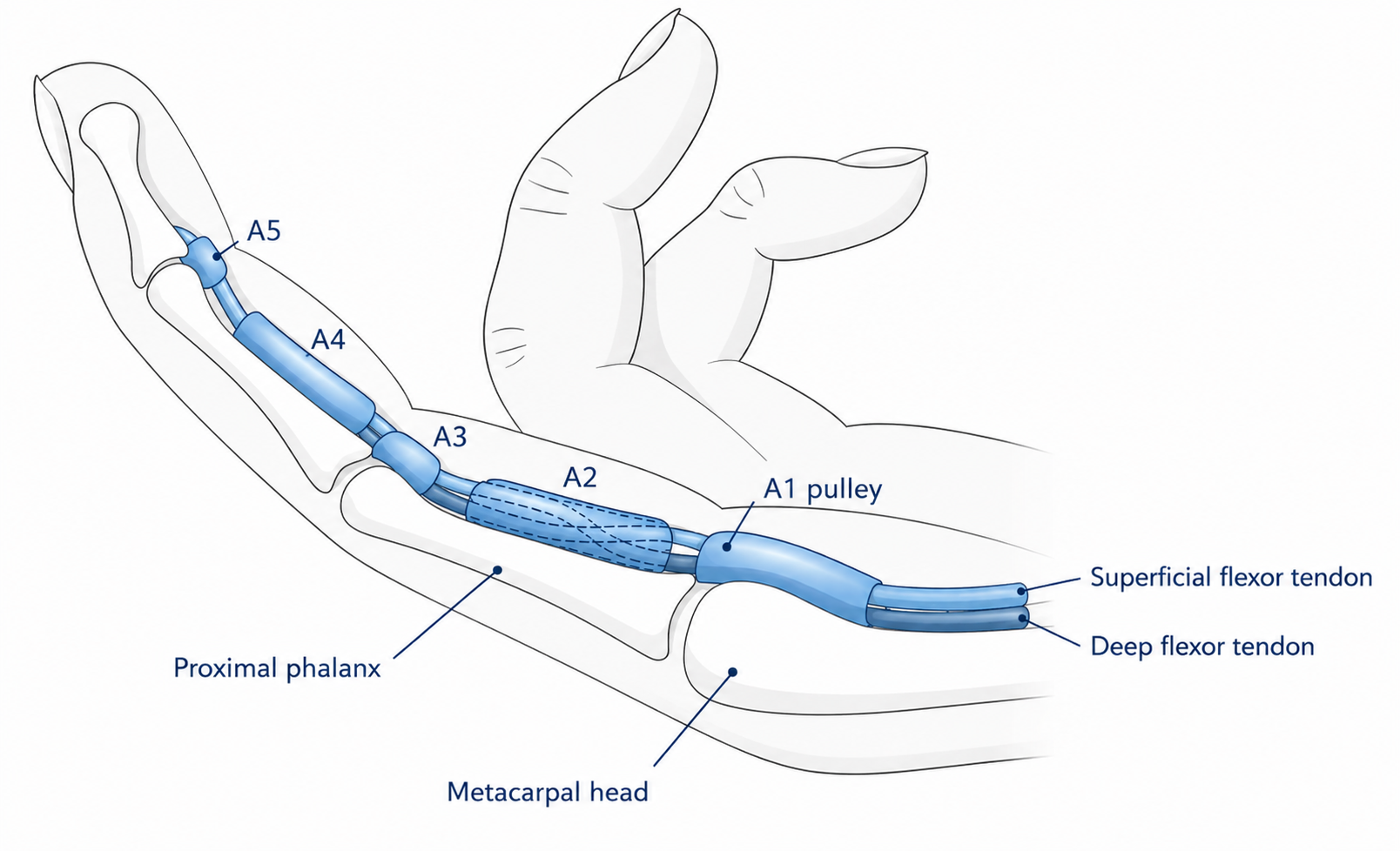
Each finger is bent by tendons that run from the forearm muscles, through the palm, and into the finger itself. These tendons do not sit loosely under the skin — they travel through a series of narrow tunnels called the flexor sheath, which runs in the palm, starting at the base of the finger, and continues to the last joint of the finger. The sheath keeps the tendon running in a straight, efficient line against the bone and, importantly, keeps it lubricated as it glides back and forth many thousands of times a day.
The entrance to this tunnel system, at the point where the finger meets the palm, is called the A1 pulley. Trigger finger develops when there is a mismatch between the width of this entrance and the tendon trying to pass through it. This can happen in one of two ways: either the tendon itself swells, most often due to inflammation of its lining, or the entrance to the tunnel narrows.
The more common cause is narrowing of the A1 pulley itself, through a process called fibrocartilaginous metaplasia. In simple terms, the pulley tissue gradually shifts from a soft, flexible type of cell (a fibroblast) to a harder, cartilage-forming cell (a chondrocyte), and loses flexibility as a result. High blood glucose is understood to accelerate this process by promoting cross-linking of collagen within the tissue, which is part of the reason trigger finger is significantly more common in people with diabetes. [1] Where inflammation of the tendon lining itself is the driver rather than pulley narrowing, this is more typical in patients with inflammatory joint conditions such as rheumatoid arthritis, systemic lupus erythematosus or gout.
Contrary to a common misconception, there is no nodule that forms on the tendon itself in adult trigger finger. What actually thickens is the A1 pulley — sometimes to as much as three times its normal thickness — narrowing the entrance the tendon must pass through. As the finger bends, the tendon is forced through this tightened entrance; as the patient tries to straighten the finger again, the tendon catches against the thickened pulley, producing the characteristic click, catch or lock. (The one genuine exception is paediatric trigger thumb, where a true nodule can form directly on the flexor tendon itself — a distinct mechanism from the adult condition.)
There is no single typical patient. Trigger finger can affect a two-year-old child born with a locked thumb — in this group, the underlying issue is usually a mismatch in tendon and tunnel size present from birth, rather than the metaplasia process seen in adults. It can affect a 20-year-old with poorly controlled insulin-dependent diabetes. Most commonly, it affects adults between roughly 40 and 60 years old, though it is also regularly seen in patients in their 80s and 90s. Diabetes, in particular, is a well-recognised risk factor across all these age groups.
In many cases, the diagnosis is straightforward: the patient demonstrates the finger clicking, catching or locking during examination. However, this is not always the case. Because the stronger flexor muscles dominate during sleep, symptoms are often at their worst first thing in the morning or during the night itself, when the finger is most likely to lock. By the time a patient reaches a clinic appointment later in the day, the finger has often loosened up and may move normally during examination.
In these situations, the more reliable sign is tenderness directly over the A1 pulley at the base of the finger, which can feel hard and nodular under the skin, sometimes with a palpable crunching sensation (crepitus) as the tendon moves beneath it.
At iiS Health, ultrasound is used as standard alongside clinical examination. This allows a direct, side-by-side comparison between a healthy tendon and the affected one: a normal tendon glides in a smooth, straight line on the scan, while an affected tendon shows visible bending or an hourglass-shaped narrowing as it is forced through the tightened entrance. This has a practical benefit beyond confirming the diagnosis — it allows the patient to see the mechanical problem directly, which makes the logic of treatment (opening up that narrowed entrance) immediately clear.
For symptoms of short duration, the first step is usually a splint worn overnight, alongside oral or topical anti-inflammatory medication.
The reasoning behind night splinting comes down to basic muscle mechanics. The muscles that bend the fingers, on the palm side of the forearm, are considerably stronger than the muscles that straighten them. While awake, a person can consciously keep the fingers extended, but during sleep this conscious control is lost, and the stronger flexor muscles win out — the fingers curl and the wrist tends to bend as well. If a finger is going to lock, it is therefore most likely to do so overnight, and spending several hours locked in a bent position tends to make the finger considerably more painful first thing in the morning. A splint that holds the finger straight overnight prevents this, and many patients notice a meaningful reduction in morning pain as a result.
Where splinting and anti-inflammatory measures are not enough, a steroid injection into the flexor tendon sheath is the standard next step — and this is where the strength of the evidence becomes genuinely important to understand, because success is far from universal.
Reported success rates for a single steroid injection vary meaningfully across studies, generally ranging from around 45% to over 90%, depending on how "success" is defined and how long patients are followed up afterwards. [2] A well-cited study following patients for six months after a single injection found a 70% rate of freedom from symptom recurrence at that point, falling to 45% by twelve months — illustrating that even a successful early response does not guarantee the problem stays resolved long-term. [3]
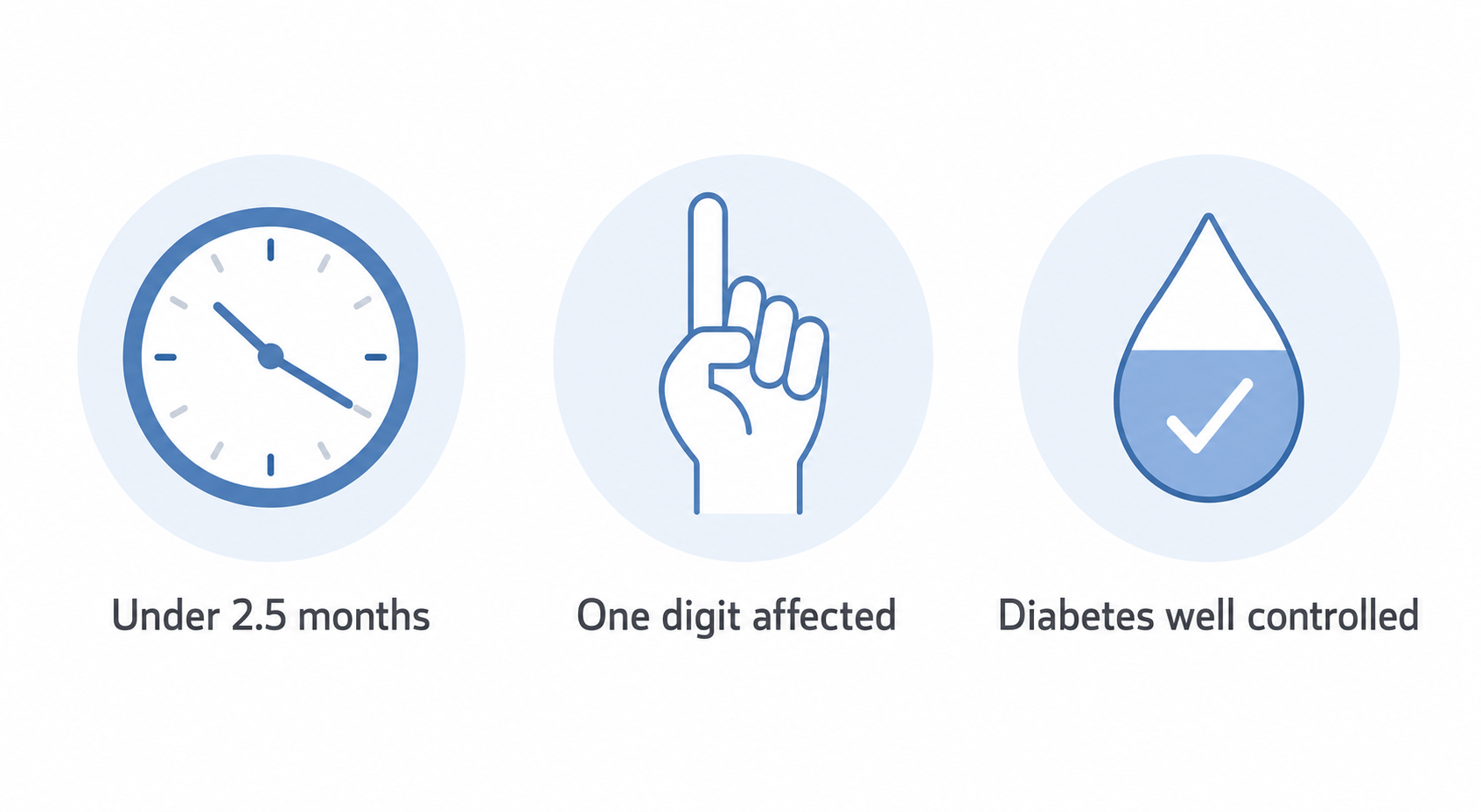
What is more clinically useful than a single headline percentage is understanding which patients are more likely to do well. The evidence consistently points to three factors:
In practice, this means the odds are best for patients with a short history, a single affected digit, and well-controlled diabetes — these patients are more likely to resolve the problem without ever needing surgery. Outside those three conditions, the odds shift the other way, and moving toward surgery sooner, rather than persisting with repeated injections, is often the more sensible course.
The mechanism by which steroid injections work is better understood than is often assumed, and it goes beyond simply damping down inflammation. Research has found that corticosteroid injection reduces the tendon sheath's production of collagen type I and proteoglycans, while increasing enzymes (MMP-1 and MMP-13) that actively break down existing collagen — meaning the injection genuinely thins the thickened pulley tissue at a biological level, rather than only masking symptoms. [7] Interestingly, the same research found that pulley thickness actually reduces more with a steroid injection than with a percutaneous (needle) release, yet the clinical outcome tends to be better with the percutaneous release — a reminder that reducing pulley thickness alone does not fully explain why some treatments succeed and others do not.
Other types of injection sometimes used elsewhere in orthopaedics — platelet-rich plasma (PRP) or hyaluronic acid, for example — are not considered appropriate treatment for trigger finger. Both are more suited to situations involving tissue injury or joint lubrication; trigger finger is neither. The problem is either a physically narrowed entrance or swollen tendon lining, and neither of these substances addresses that mechanism directly.
Tendon gliding exercises — bending and straightening the finger through its full range, and isolating one flexor tendon at a time so it glides independently of the other — are frequently recommended, usually by a hand therapist. The A1 pulley itself is a static structure and does not move; the logic behind these exercises is that repeatedly gliding the tendon through it keeps stretching the pulley, which may help slow how quickly it narrows.
It is worth being straightforward about the evidence here rather than overstating it. A 2025 randomised controlled trial specifically testing finger gliding exercises alongside steroid injection found no statistically significant additional clinical benefit compared with injection alone. [5] This does not mean the exercises are unhelpful or should be abandoned — the underlying physiological rationale is sound, and hand therapists continue to recommend them as part of a broader recovery plan — but it is more accurate to describe them as a reasonable, low-risk adjunct than a proven independent treatment in their own right.
Several factors reliably shift the decision toward surgery rather than continuing with injections:
Trigger finger release is a short day-case procedure, and at iiS Health it is offered in two forms.
Open release involves a small incision in the palm to divide the tightened A1 pulley directly under vision. Because the procedure is performed under local anaesthetic with the patient awake, the result is immediately obvious — the patient can move the finger on the table and confirm the clicking and locking have resolved before leaving.
Ultrasound-guided release achieves the same result — dividing the A1 pulley — through a small needle hole rather than an open incision, using real-time ultrasound imaging to guide the release accurately. The main advantage is a considerably faster return to normal activity, since there is no surgical wound to protect.
One anatomical detail worth being aware of: a smaller, less consistently developed pulley just towards the wrist side of the A1 pulley, sometimes called the A0 pulley, occasionally contributes to triggering as well. Because it is less well defined, it is not always clearly visible on ultrasound, which means a small minority of patients treated with ultrasound-guided release may find symptoms persist and require a further, open procedure to address it directly.
Recovery differs meaningfully between the two techniques.
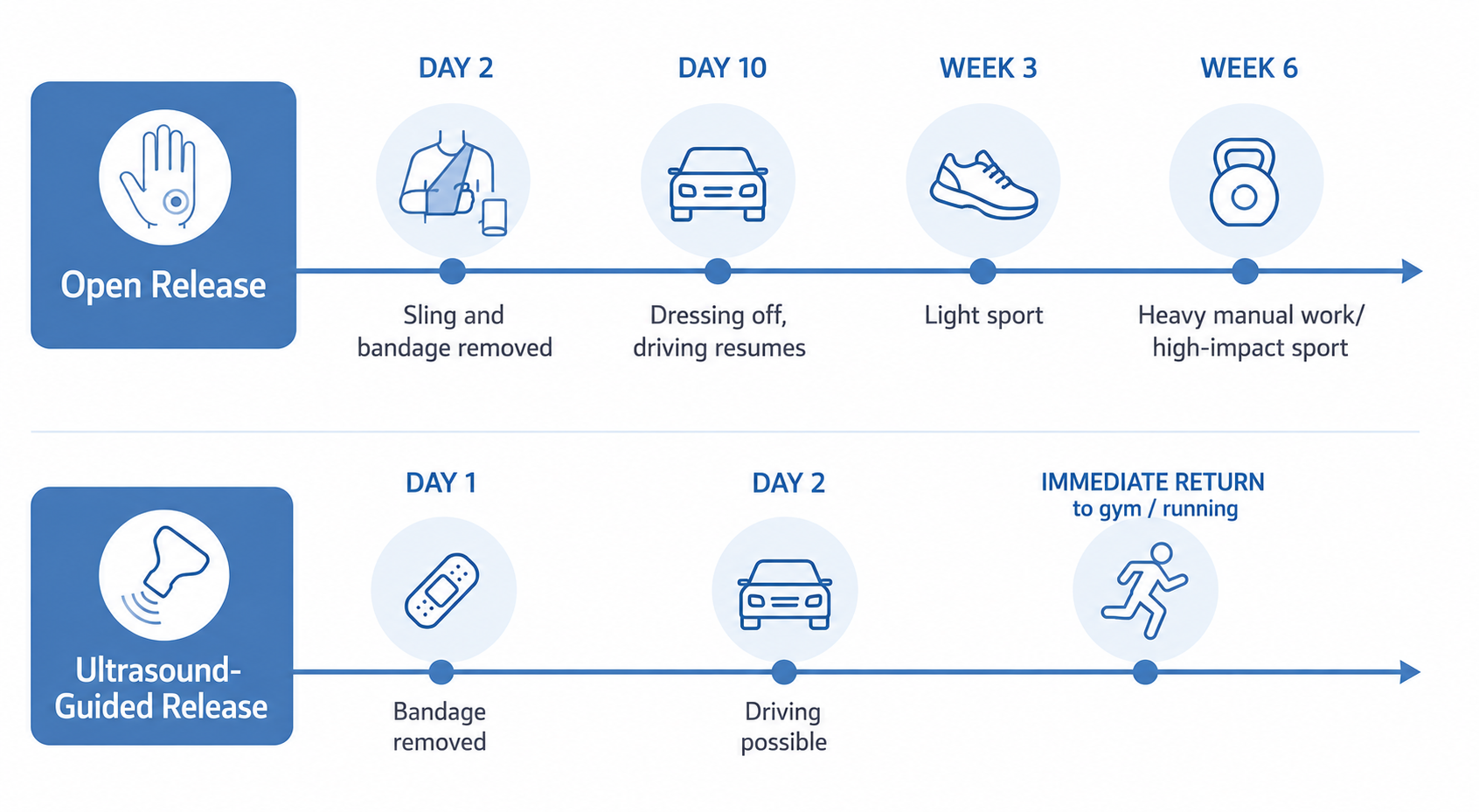
After open release: a padded dressing and sling are worn for the first 48 hours, after which a smaller adhesive dressing remains in place for a total of around ten days while the wound heals — longer than some other surgical specialties recommend, reflecting the fact that hands are in near-constant contact with surfaces of every kind. Driving typically resumes once the wound is uncovered, at around ten days. Return to desk-based work is usually possible within a couple of days; light manual work, around four weeks; heavy manual work, around six weeks. For sport, the general principle is that nothing is damaged by returning early once the wound has healed — it is a question of comfort rather than risk — with most racket sports and swimming manageable from around three weeks, and higher-impact activity such as rugby closer to six weeks.
After ultrasound-guided release: the initial bandage comes off after 24 hours, with no ongoing wound to protect. Driving is often possible from around 48 hours if the hand feels strong enough. Return to the gym or running can begin almost immediately. This faster recovery is a genuine practical advantage for patients with time-sensitive commitments — one example is a marathon runner treated by ultrasound-guided release specifically to avoid ten days out of training ahead of race day, who was running again the following day.
As with any procedure, trigger finger release carries some risk, though serious complications are uncommon:
Where a tourniquet would traditionally be used to keep the surgical field clear of blood, many clinics — iiS Health included — instead use a WALANT technique (local anaesthetic combined with adrenaline), which achieves a bloodless field without the discomfort of a tourniquet. Ultrasound-guided release does not require a tourniquet at all.
"Trigger finger and Dupuytren's contracture are the same thing."
They are not, though the confusion is understandable, since both can affect the same fingers. The distinguishing feature is that a finger affected by trigger finger, even when locked, can be straightened — with effort, or with help from the other hand — whereas a finger affected by Dupuytren's contracture cannot be passively straightened at all, because the underlying problem is thickened tissue in the palm's fascia rather than a tendon catching on a pulley.
Interestingly, the two conditions are more closely linked than their different mechanisms might suggest. A 2019 study found a statistically significant association between trigger finger and Dupuytren's contracture that was not explained by their shared risk factors, such as diabetes, smoking or manual labour — patients with one condition appear to be at meaningfully increased risk of the other. [6] The exact biological reason for this link is not yet fully established.
"If you have trigger finger, you will eventually need surgery."
This is not the case. Many patients respond fully to splinting, anti-inflammatory measures or a single injection, and some patients' symptoms settle on their own without ever needing formal treatment.
Persistent clicking, catching or locking in a finger or thumb — particularly if it is affecting grip, work, or sleep — is a reasonable prompt to seek a specialist assessment rather than waiting to see if it resolves on its own, especially given how strongly the evidence points to earlier treatment producing better outcomes. iiS Health offers on-site ultrasound assessment as standard, allowing diagnosis and a clear treatment plan in a single visit.








.png)